Forearm Atrophy and Denervation on MRI
Hi all, here a little video I just did for my students in the Virtual MSK Fellowship. the edema pattern shows median nerve involvement, so AIN syndrome is a good DDx. the edema in the Pronator teres might be also DOMS related as a frequent gym goer. a relevant pathology of the nerve is not seen. here some resources: https://radsource.us/median-nerve-entrapment/ https://radiopaedia.org/articles/anterior-interosseous-nerve-syndrome-1 This type of detailed case analysis is exclusive to my MSK Fellowship members. They get to submit their own challenging cases and receive personalized video feedback like this, helping them master complex diagnoses and become the "Go-To MSK Expert" in their practice. Ready to join a community of dedicated radiologists and elevate your MSK skills? Book a call to learn more: www.onlinemskfellowship.com (watch or click on case study - to book the call).
2
0

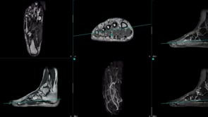
example plantar vein thrombosis
plantar vein thrombosis can be hard to see if not expected or never seen before. https://vimeo.com/1021589562/65cf61a363?share=copy https://www.youtube.com/watch?v=OK0_MTI27vc
13
5
New comment 2d ago

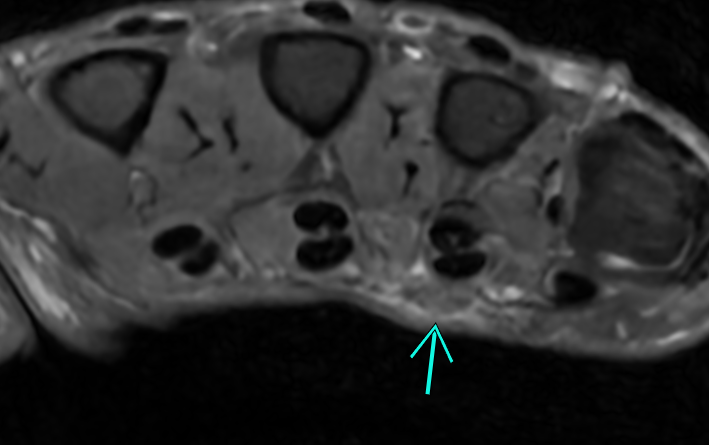
Duyputren's disease on MRI
here an example of early duyputrens disease on MRI with pretendinous cords. Can be hard to see. here a great article with amazing illustrations! https://radsource.us/dupuytrens-disease/ have you seen this before on MRI?
5
3
New comment 2d ago

3rd head of gastrocnemius muscle
here a lovely anatomic variant of a 3rd head of the gastrocnemius muscle. https://radiopaedia.org/articles/third-head-of-gastrocnemius From radiopaedia: Third head of gastrocnemius, also known as accessory head of gastrocnemius, is a normal anatomical variation where there is an accessory muscle bundle of the gastrocnemius muscle in addition to the medial and lateral heads. Epidemiology It is the most common variation of the gastrocnemius. Its occurrence was estimated to be between 2.0% and 5.5% of the normal population 1,4. Clinical picture Most of the cases are asymptomatic. It can cause compression upon popliteal vessels resulting in clinical symptoms such as popliteal artery entrapment syndrome resulting in claudication in young patients and popliteal vein entrapment 2. Rarely, it can cause sural nerve entrapment 5. Radiographic features It is usually seen in cross-sectional imaging modalities and best identified with MRI. It arises near midline of posterior distal femur and joins the medial or lateral head of gastrocnemius muscle, coursing lateral to the popliteal vessels. The size varied from a thin threadlike muscle to a rather bulky muscle. History and etymology It was first described in 1813 by Kelch 3 and was further detailed by Frey in 1919 4.
5
2
New comment 11d ago

Don't miss this!
Here is an interesting case I had today. Come to the MSK school (free!) and watch the video. -Chris
10
4
New comment 14d ago

1-30 of 46

skool.com/mskrad
We learn MSK radiology together as a community! Grab Your FREE 5-Minute Knee MRI Masterclass today. This is a limited-time offer.
powered by


